tree in bud radiology assistant
We Cover Your Travel Housing Credentialing Privileging Malpractice. Diagnosis Pathophys Radiology Pulmonary CTChest TreeInBud Diagram RadiologyAssistant.

Scielo Brasil Padroes Tomograficos Das Doencas Intersticiais Pulmonares Difusas Com Correlacao Clinica E Patologica Padroes Tomograficos Das Doencas Intersticiais Pulmonares Difusas Com Correlacao Clinica E Patologica
One characteristic feature of bronchiolar disease is a tree-in-bud pattern on computed tomography CT.
. 2 Aquino SL Gamsu G Webb WR Kee ST. The tree-in-bud distribution is often. Tree-in-bud almost always indicates the presence of.
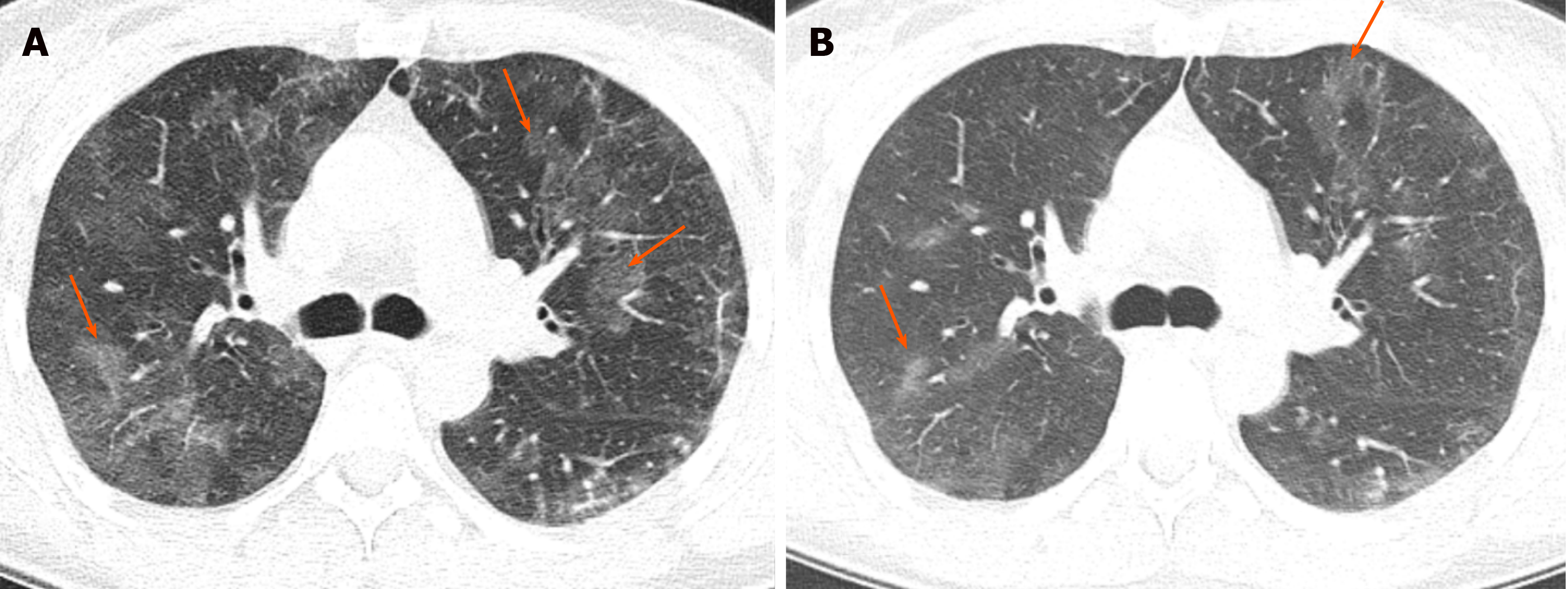
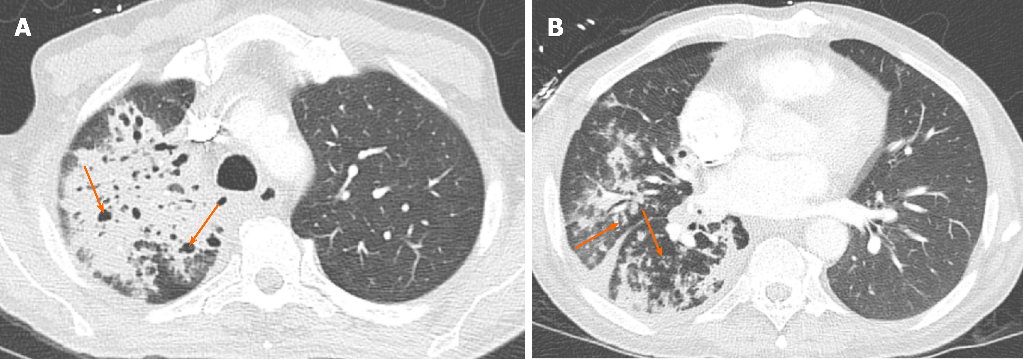
Areas of consolidation along with ground glass opacity involving the lingual contiguous with the inferior lateral portion of the left upper lobe abutting the left major fissure. The Radiology Assistant Basic Interpretation Idiopathic Pulmonary Fibrosis Pulmonary Fibrosis Langerhans Cell Histiocytosis. To describe the appearance of the endobronchial spread of mycobacterial tuberculosis.
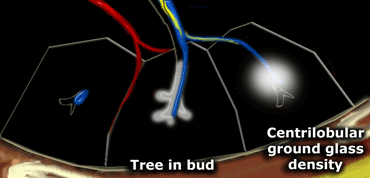
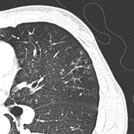
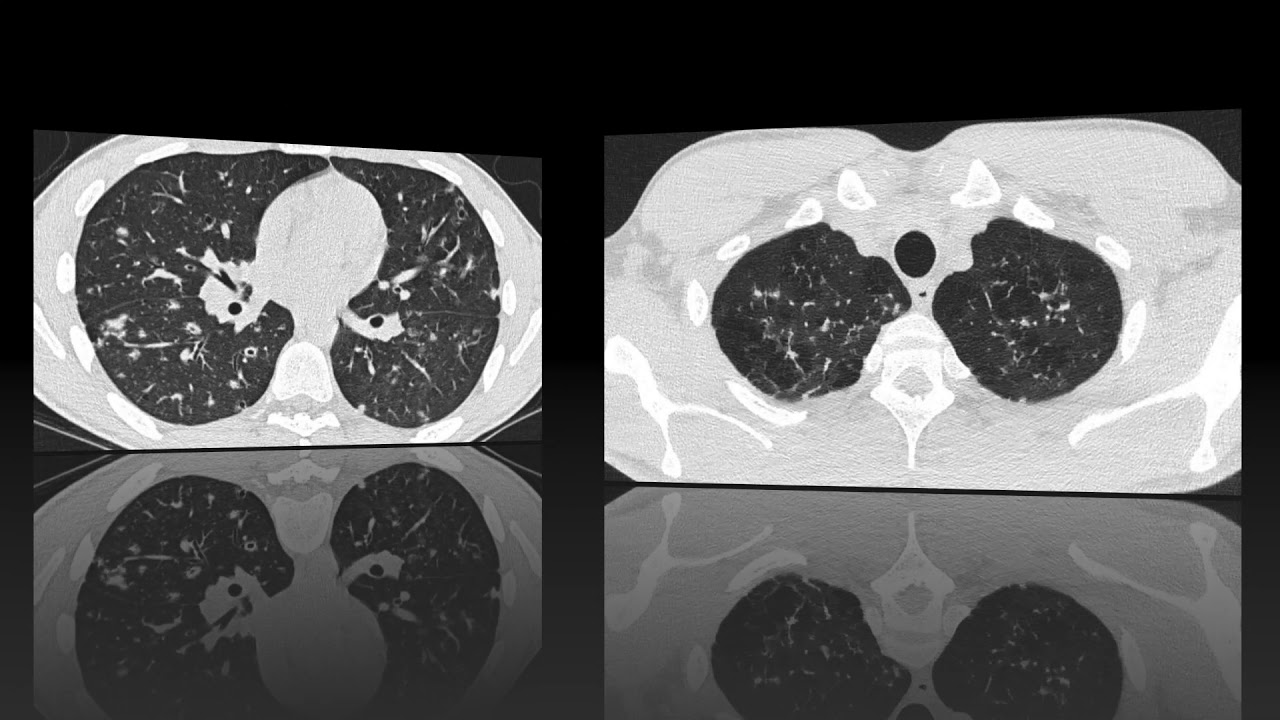
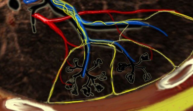
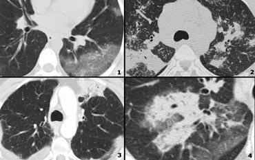
It represents dilated and impacted mucus or pus-filled centrilobular bronchioles. Tree-in-bud describes the appearance of an irregular and often nodular branching structure most easily identified in the lung periphery. 1-4Reported causes include infections aspiration and a variety of infl ammatory.
Abnormal tree-in-bud bronchioles can be distinguished from normal centrilobular bronchioles by their more irregular appearance lack of tapering or knobbybulbous appearance at the tip of their branches. Tree-in-bud TIB opacities are a common imaging finding on thoracic CT scan. Job Listings From Thousands of Websites in One Simple Search.
Fig 7 Tree In Bud Sign Chest Ct Shows Tree In Bud Images Schematic Drawings And Corresponding Picture Refe Radiology Radiology Imaging Medical Radiography. However vascular lesions involving the arterioles and capillaries may simulate the centrilobular small nodules and. Ad Enter Your ZIP Find Jobs Now Hiring Near You.
Pus mucus or inflammatory exudate centrilobular bronchioles. Crossref Medline Google Scholar. Ad Comprehensive 247 Support for Radiology Jobs Is Just a Phone Call Away.
Other causes could be immunological congenital and idiopathic disorders as well as aspiration or inhalation of. A similar pattern but smaller areas are identified involving the lateral segment middle lobe. Tree in bud radiology assistant Monday April 4 2022 Edit.
The list of the most frequent differential diagnoses for tree-in-bud sign includes infections with Mycobacterium tuberculosis nontuberculous mycobacteria and other bacterial fungal or viral pathogens. The tree-in-bud sign is a common finding in HRCT scans. Tree in bud opacification refers to a sign on chest CT where small centrilobular nodules and corresponding small branches simulate the appearance of the end of a branch belonging to a tree that is in bud.
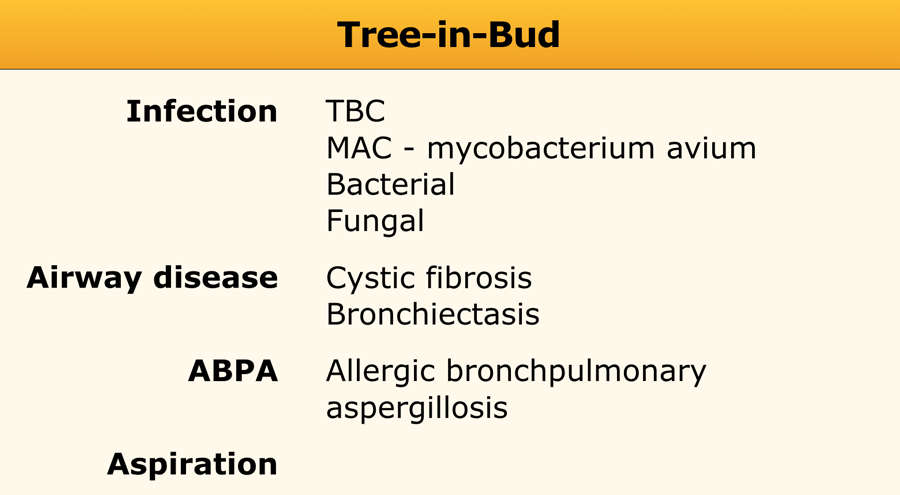
Frequency and significance on thin section CT. Endobronchial spread of infection TB MAC any bacterial bronchopneumonia Airway disease associated with infection cystic fibrosis bronchiectasis less often an airway disease associated primarily with mucus retention allergic bronchopulmonary aspergillosis asthma. J Comput Assist Tomogr 1996.
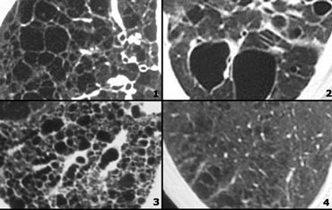
J Comput Assist Tomogr 1996. Identification and evaluation of centrilobular opacities on high-resolution CT. The small nodules represent lesions involving the small airways.
Its microbiologic significance has not been systematically evaluated. Tree-in-bud sign refers to the condition in which small centrilobular nodules less than 10 mm in diameter are associated with centrilobular branching nodular structures 1 Fig. Tree-in-bud pattern simulating diffuse panbronchiolitis but without cylindrical bronchiectasis.
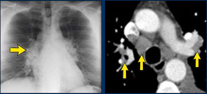
There is a cavitating lesion and typical tree-in-bud appearance. Diagnosis Pathophys Radiology Pulmonary CTChest TreeInBud Diagram RadiologyAssistant. The other is centrilobular nodules.
The tree-in-bud pattern was first used as a descriptor by Im et al. This pattern is most pronounced in the lung periphery and is usually associated with abnormalities of the larger. Originally and still often thought to be specific to endobronchial Tb the sign is actually non-specific and is the manifestation of pus mucus fluid or other.
The Tree-in-Bud Pattern. Tree-in-bud TIB is a radiologic pattern seen on high-resolution chest CT reflecting bronchiolar mucoid impaction occasionally with additional involvement of adjacent alveoli. With Snagajob you can apply to jobs in minutes and easily search for your right-fit.
All jobs Find your new job today. 3 Gruden JF Webb WR. Multiple centrilobular nodules many with a tree in bud.
Tree-in-bud appearance represents dilated and fluid-filled ie.
The Radiology Assistant Hrct Basic Interpretation
The Radiology Assistant Hrct Basic Interpretation
Chronic Airspace Disease Review Of The Causes And Key Computed Tomography Findings
The Radiology Assistant Hrct Basic Interpretation
Tree In Bud Sign Lung Radiology Reference Article Radiopaedia Org

Tree In Bud Sign Lung Radiology Reference Article Radiopaedia Org

Pin On Chest Ct Mri
The Radiology Assistant Hrct Common Diagnoses
2

Learningradiology Lung Abscess Pulmonary Lunges Pulmonary X Ray
The Radiology Assistant Hrct Basic Interpretation

Cavity Consolidation With Multiple Areas Of Nodular Opacity Showing Tree In Bud Appearance Most Likely Possibility Of E Opacity Abstract Artwork Abstract

Palla S Sign
2
The Radiology Assistant Hrct Common Diagnoses
The Radiology Assistant Hrct Basic Interpretation
Chronic Airspace Disease Review Of The Causes And Key Computed Tomography Findings

On The Left Some Diseases With A Nodular Pattern 1 Grepmed

The Radiology Assistant Lung Hrct Basic Interpretation Radiology Imaging Radiology Diagnostic Imaging